Bilateral nevus of Ota in a light-skinned woman
Published Web Location
https://doi.org/10.5070/D34fg002sdMain Content
Bilateral nevus of Ota in a light-skinned woman
Bojana Jovovic-Dagovic MD, Ana Ravic-Nikolic MD, Vesna Milicic MD, Gordana Ristic MD
Dermatology Online Journal 13 (3): 19
Department of Dermatology, Clinical Center Kragujevac, Kragujevac, Serbia Abstract
Nevus of Ota is a dermal melanocytosis, clinically localized on skin that is innervated by the first and second branches of the trigeminal nerve. It occurs almost entirely in Asian people. This manifestation is rarely described in light-skinned non-Asian persons. We present a case of bilateral Ota nevus in a 47-year-old light-skinned non-Asian woman.
Clinical synopsis
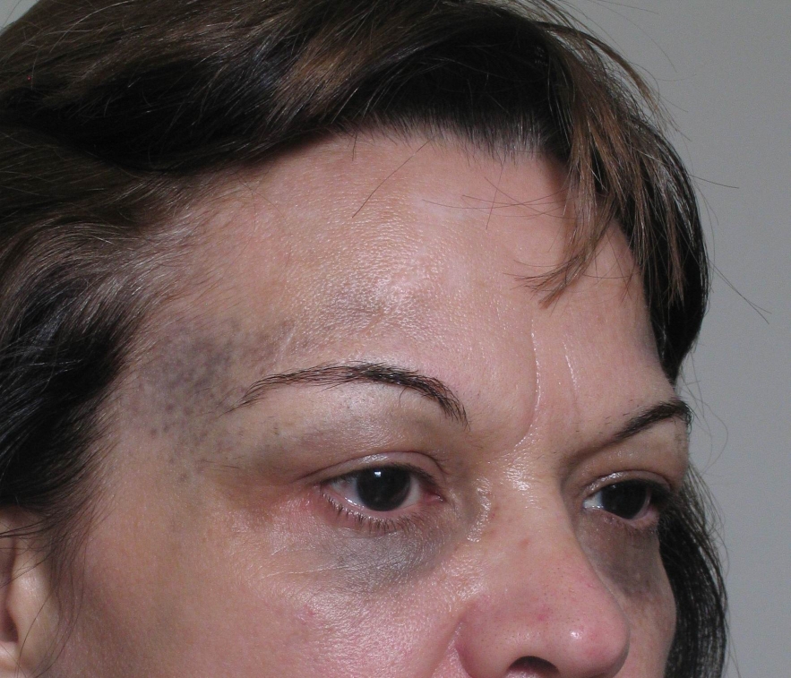 | 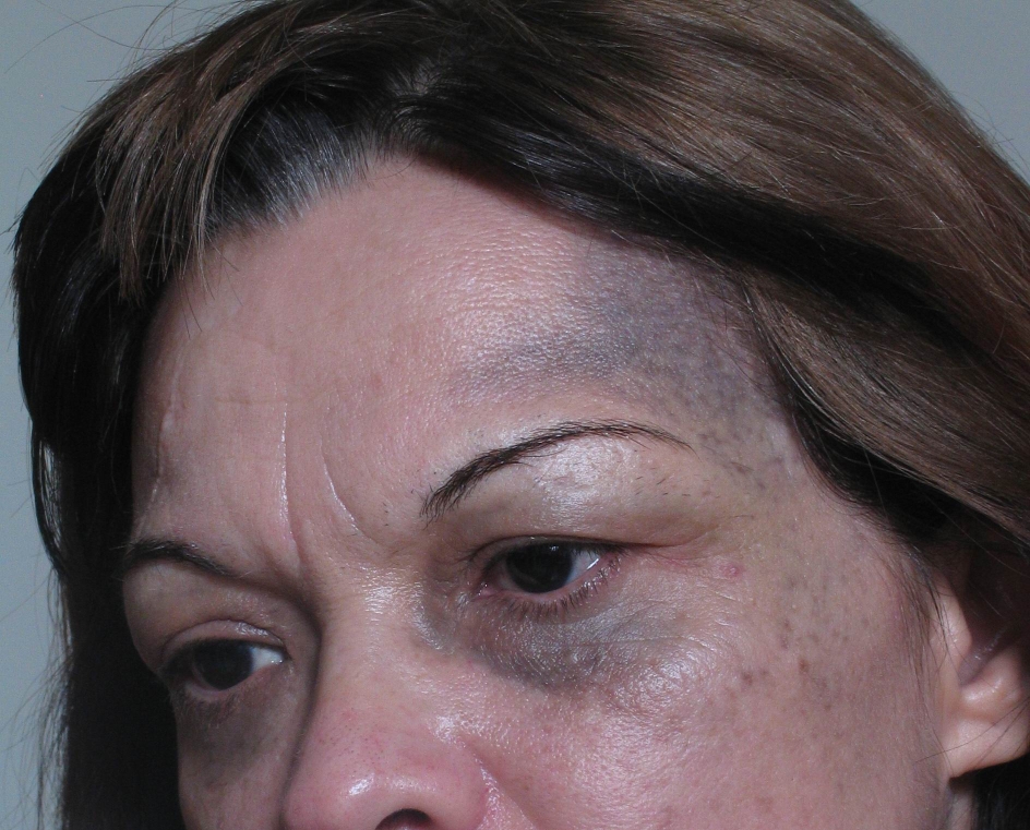 |
| Figure 1 | Figure 2 |
|---|
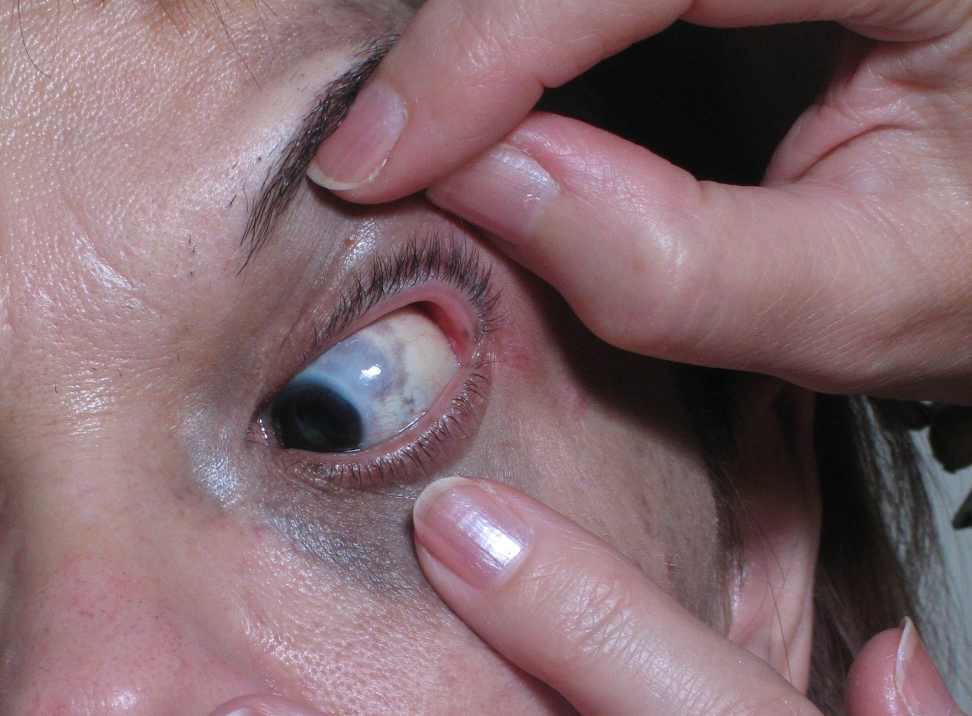 | 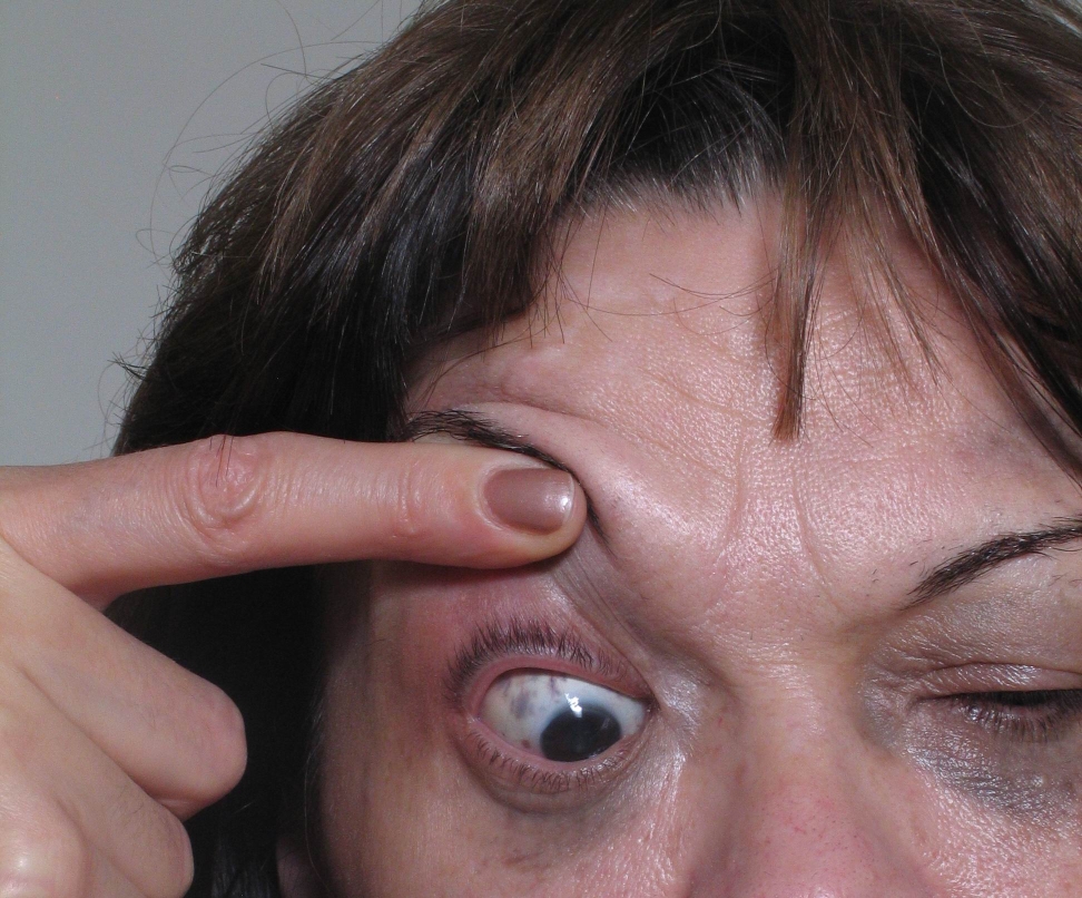 |
| Figure 3 | Figure 4 |
|---|
A 47-year-old light-skinned non-Asian woman presented with bilateral manifestations of nevus of Ota. The ocular hyperpigmentation was present at birth and the skin lesions appeared in early childhood. Clinically, flat blue-gray pigmentation with an irregular border was present on the skin of the lateral ocular areas. Bilaterally, the sclerae were also involved. Nasal and oral mucosa were not affected. Ophthalmologic examination showed no abnormalities. Audiometric examination was within normal limits.
Comments
Nevus of Ota was first described by a Japanese dermatologist, M.T. Ota, in 1939 [1]. Histopathology of affected skin shows the presence of dendritic cells containing melanin in the dermis [2]. Ota nevus can be congenital or acquired in adolescence. It occurs almost entirely in persons of Asian descent. Manifestations in fair-skinned non-Asians are very rare [3]. The clinical manifestations are usually unilateral; only 5 percent of cases are bilateral. Clinically, blue-gray macular pigmentation with irregular border involves skin that is innervated by the first and second branches of the trigeminal nerve. Extrancutaneous manifestations include ocular involvement of sclera, episclera, conjunctiva, cornea, retina, and uveal tract. Similar discoloration can be observed in oral mucosa (buccal and palatal), as well as in nasal mucosa and the tympanic membrane. Leptomeninges can also be affected. The occurrence of melanoma has been rarely described [4].
Various therapies have been used successfully. Cosmetic coverup products can be used for camouflage. Cryosurgery and microsurgical treatments can leave disfiguring scars and are not recommended. Chemical bleaching agents can damage epidermal melanocytes and provoke permanent hypopigmentation or depigmentation [5]. Combined dermabrasion and the carbon dioxide snow method has produced good results [6].
In recent years the use of laser therapy has been very effective and gives new hope for patients with nevus of Ota [5, 7]. Various lasers are used in treatment of pigmentary disorders and the best results for treatment of this condition appears to be obtained with Q-switched Nd-YAG, ruby and alexandrite lasers. The long wavelengths penetrate deeply into the dermis and provoke selective photothermal and photomechanical destruction of dermal melanocytes [8]. After irradiation, the shape of dermal melanocytes and their melanosomes is changed. Melanosomes are fragmented into smaller pieces and their cell membranes are disrupted; the nucleus is fragmented or destroyed. Destruction of dermal melanocytes can be achieved without collateral injury to the surrounding tissue [9]. There is no significant epidermal pigmentation in patients with nevus of Ota. Melanosomes in epidermal melanocytes are different compared to dermal melanocytes; they are smaller and more numerous. After laser therapy epidermal melanocytes are reversibly changed; light microscopy shows expansion of extracellular space, swollen mitochondria, dilatation of endoplasmic reticulum, and vacuolated melanosomes. One year after the treatment normal cell structure is completely restored [10].
Complications such as mild purpura, erythema and edema can follow the treatment and improves after a few days. The most frequent complication is hypopigmentation, which can be transient or permanent. Hyperpigmentation can also occur 2-3 weeks after treatment and lasts for a few months. Topical tretinoin gel, hydroquinone, and corticosteroid creams can be used in the treatment of post-inflammatory hyperpigmentation [8, 11, 12]. Laser therapy is very effective in the treatment of nevus of Ota, and recurrence is rare [5, 7, 8, 9].
References
1. Ota M. Nevus fuscocoerulens ophtalmomaxillaris. Tokio Med J. 1939; 63: 1243-1245.2. Kishikawa T, Suzuki T, Sasakia Y, Aihara K, Hirayama T. Characterization of melanosomes and melanogenesis in cells cultured from Ota's nevus. J Submicrosc Cytol Pathol. 1997 Jul; 29(3):339-352. PubMed
3. Turnbull JR, Assaf Ch, Zouboulis C, Tebbe B. Bilateral naevus of Ota: a rare manifestation in a Caucasian. J Eur Acad Dermatol Venereol. 2004 May; 18(3): 353-355. PubMed
4. Patel BC, Egan CA, Lucius RW et al. Cutaneous malignant melanoma and oculodermal melanocytosis (naevus of Ota): report of a case and review of the literature. J Am Acad Dermatol. 1998; 38:862-865. PubMed
5. Yang HY, Lee CW, Ro YS, Yu HJ, Kim YT, Kim JH, Kim JH. Q-switched ruby laser in treatment of nevus of Ota. J Korean Med Sci. 1996Apr; 11(2):165-170. PubMed .
6. Hata Y, Matsuka K, Ito O, et al. Treatment of naevus of Ota: combined skin abrasion and carbon dioxide snow method. Plast Reconstr Surg. 1996; 97: 544-554. PubMed .
7. Kang W., Lee E., Choi GS. Treatment of Ota's nevus by Q-switched alexandrite laser: therapeutic outcome in relation to clinical and histopathological findings. Eur J Dermatol. 1999 Dec; 9(8): 639-643. PubMed
8. Omprakash N. Treatment of nevus Ota by Q-switched, frequency doubled, ND: YAG laser. Indian J Dermatol Venereol Leprol 2002; 68: 94-95.
9. Lu Z., Chen J., Wang X., Fang L., Jiao S., Huang W. Effect of Q-switched alexandrite laser irradiation on dermal melanocytes of nevus of Ota. Chin Med J 2000; ll3 (1):49-52. PubMed
10. Lu Z., Chen J., Wang X., Fang L., Jiao S., Huang W. Effect of Q-switched Alexandrite laser irradiation on epidermal melanocytes in treatment of nevus Ota. Chin Med J 2003 Apr: ll6 (4):597-601. PubMed
11. Chan HH., Leung RS., Ying SY., Lai CF., Kono T., Chua JK., Ho WS. A retrospective analysis of complications in the treatment of nevus of Ota with the Q-switched Alexandrite and Q- switched Nd: YAG lasers. Dermatol Surg. 2000 Nov; 26 (11): 1000-1006. PubMed
12. Kono T., Nozaki M., Chan HH., Mikashima Y. A retrospective study looking at the long-term complications of Q -switched ruby laser in the treatment of nevus of Ota. Lasers Surg Med. 2001; 29(2): 156-159. PubMed
© 2007 Dermatology Online Journal