Erysipelas of the left upper limb occurring after elbow dislocation
Published Web Location
https://doi.org/10.5070/D38w94q2sdMain Content
Erysipelas of the left upper limb occurring after elbow dislocation
Talel Badri MD, Mourad Mokni MD, Maha Ben Sassi MD, Faika Cherif MD, Mohamed Iyadh Azaiz MD, Amel Ben Osman Dhahri MD
Dermatology Online Journal 12 (4): 9
Dermatology Department, La Rabta Hospital, Rue Jabbari, 1007 Tunis- Tunisia. mourad.mokni@rns.tnAbstract
BACKGROUND: Erysipelas is an acute infection occurring chiefly in the lower limbs, rarely in the upper limbs. OBSERVATION: A 45-year-old patient suffering from Charcot-Marie-Tooth disease with neuropathy of the limbs, presented with fever and a 24-hour history of a well-circumscribed inflammatory and infiltrated plaque of the left arm. Erysipelas was diagnosed and intravenous penicillin was administered leading to regression of the inflammatory signs, however edema persisted in the inner part of the left elbow. An x-ray showed left elbow dislocation. The patient revealed trauma of the left upper limb 5 weeks before. DISCUSSION: The occurrence of erysipelas is usually associated with lymphatic edema or venous incontinence. Lymphatic lesions due to radiotherapy or surgery may afflict draining vessels leading to venous and lymphatic stasis and then infection occurs. We find no reported cases of erysipelas following elbow dislocation but we postulate its pathogenesis to be similar.
Erysipelas is an acute infectious disease occurring chiefly in the lower limbs. It involves the dermis and dermal lymphatics as opposed to cellulitis which involves the deep dermis and subcutaneous tissue. The occurrence of erysipelas in the upper limbs is rare.
Clinical synopsis
We report the case of a 45-year-old woman suffering since childhood from Charcot-Marie-Tooth disease Type 1 of uncertain inheritance mode. For the previous 5 years she suffered from slowly progressive neuropathic disorders, including anesthesia of the feet and hypesthesia of the legs and upper limbs. Neuromuscular biopsy revealed plurisegmental demyelinization consistent with a severe axonal neuropathy.
The patient presented with fever, fatigue, and a warm erythematous plaque of the left upper limb that occurred and developed rapidly within 24 hours. On physical examination, the patient was febrile (38.6° C) and had a well-circumscribed erythematous, edematous and inflammatory plaque with distinct edge on the left arm and forearm, within which were multiple blisters containing serosanguinous fluid (Fig. 1).
Bilateral palmar scratches were also observed but no associated nodule was found. Mobilization of the left elbow was slightly painful and neurological examination confirmed hypesthesia and absence of tendon reflexes in the limbs. Biological tests showed leukocytosis at 13x106/ml. Cultures of fluid from two blisters were negative. Arterio-venous Doppler examination failed to show vascular thrombosis. Erysipelas of the left upper limb was thus diagnosed and penicillin-G therapy was administered at the dose of 12 million IU per day intravenously for 15 days leading to the regression of the inflammatory plaque; however, there was persistent edema of the inner face of the left elbow as well as a functional impairment. On further investigation, the patient revealed a neglected trauma of the left upper limb 5 weeks prior. X-ray examination showed left elbow dislocation.
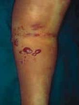 | 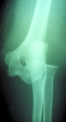 |
| Figure 1 | Figure 2 |
|---|---|
| Figure 1. Edematous and inflammatory sheet of the left upper limb Figure 2. X-ray showing elbow dislocation | |
The patient was referred to orthopedics and treated with external fixation. No recurrence was noted within a 5-year followup.
Discussion
Erysipelas is usually seen in the lower limbs and less commonly in the face or the upper limbs. This latter location represents 1-9 percent of erysipelas sites [1]. The major erysipelas organism is Streptococcus pyogenes. Risk factors for erysipelas occurrence are controversial. There is only a case-control study that analyzed different risk factors for erysipelas of the leg [2]. According to the authors, the main risk factors are: lymphatic edema, the existence of a site of entry, lower limb edema, venous insufficiency, and overweight. Erysipelas of the upper limbs is chiefly seen in women treated by radiotherapy for breast cancer, with or without lymphadenectomy [1, 3]. The resulting lymphatic drainage disorder leads to lymphatic and venous stasis, which is a favorable condition for infection.
Studer-Sachsenberg et al. [4] reported cases of cellulitis of the hip in patients carrying prosthesis after hip surgery. These cases occurred several weeks to several years after surgery, which eliminates in principle prosthesis infection. The pathogenetic mechanism may be related to lymph vessel damage occurring during surgery leading to venous and lymphatic stasis. Erysipelas and cellulitis of the leg occurring after venectomy for coronary-artery bypass are reported by several authors [5, 6]. Venous and lymphatic lesions are noted by Baddor et al. [6]. To our knowledge, no cases of erysipelas following elbow dislocation are reported. The pathogenetic mechanism might be similar to that described in surgical trauma. In our patient, erysipelas was diagnosed because of the following findings: acute onset of fever associated with a well-circumcized inflammatory plaque of the left upper limb with a distinct edge, a presumptive site of entry—palmar scratches, and leucocytosis. Bacteriological study, as for our case, is often not helpful for the diagnosis because of a low sensitivity (5-41 % positive [7]).
Charcot-Marie-Tooth disease is a hereditary neurological disease with a slow evolution. In the limbs, it occurs as amyotrophy and a loss of sensitivity [8] as observed in our patient. Type 1 usually starts in adolescence or early adulthood. Clinical signs are progressive weakness of distal limb muscles initially involving the legs and later, the hands, the arms, the forearms. Sensitive anomalies occur late in the disease. Several cases of type 1 are attributed to de novo mutations and appear sporadically [9].
In our patient having sensitivity disorders, elbow dislocation was initially misdiagnosed because of the lack of pain. Furthermore, elbow deformity was masked by the important edema of the limb. Antibiotics were followed by apyrexia and regression of inflammation. Residual functional impairment led to the x-ray examination that revealed elbow dislocation.
References
1. Becq-Giraudon B. L’érysipèle : prévention primaire et secondaire. Ann Dermatol Venereol 2001;128:368-75. PubMed2. Dupuy A, Benchikhi H, Roujeau JC, Bernard P, Vaillant L, Chosidow O, Sassolas B, Guillaume JC, Grobb JJ, Bastuji-Garin S. Risk factors for erysipelas of the leg (cellulitis): case-control study. Br Med J 1999;318:1591-4. PubMed
3. Ben Salah H, Siala W, Maaloul I, Bouzid F, Frikha M, Daoud J. Erysipelas after breast cancer treatment. Tunis Med 2002;80:465-8. PubMed
4. Studer-Sachsenberg EM, Ruffieux P, Saurat JH. Cellulitis after hip surgery: long term follow up of seven cases. Br J Dermatol 1997;137:133-6. PubMed
5. Dan M, Heller K, Shapira I, Vidne B, Shibolet S. Incidence of erysipelas following venectomy for coronary artery bypass surgery. Infection 1987;15:107-8. PubMed
6. Baddour LM, Bisno AL. Non-group A beta hemolytic streptococcal cellulitis. Association with venous and lymphatic compromise. Am J Med 1985;79:155-9. PubMed
7. Vaillant L. Critères diagnostics de l’érysipèle. Ann Dermatol Venereol 2001;128:326-33. PubMed
8. Rosa A. Maladies hérédodégénératives du système nerveux. In: Godeau P, Piette JC, Herson S, eds. Traité de médecine, 2nd edition. Paris:Flammarion, 1992;3101-6.
9. Bertorini T, Narayanaswami P, Rashed H. Charcot-Marie-Tooth disease (hereditary motor sensory neuropathies) and hereditary sensory and autonomic neuropathies. Neurologist 2004;10:327-37. PubMed
© 2006 Dermatology Online Journal