Cutaneous larva migrans in an unusual site
Published Web Location
https://doi.org/10.5070/D32fb6q1ckMain Content
Cutaneous larva migrans in an unusual site
SK Malhotra, Rakesh T Raj, Manjeet Pal, Vippan Goyal, Shweta Sethi
Dermatology Online Journal 12 (2): 11
G.G.S. Medical College, Faridkot-151203, Punjab, India. manjeet_pal@yahoo.comIntroduction
Cutaneous larva migrans is a common tropically-acquired cutaneous eruption. It presents as an erythematous, serpiginous, pruritic, cutaneous eruption associated with percutaneous penetration and subsequent migration of larvae of various nematode parasites [1]. We report a case of cutaneous larva migrans involving the anterior abdominal wall.
Clinical synopsis
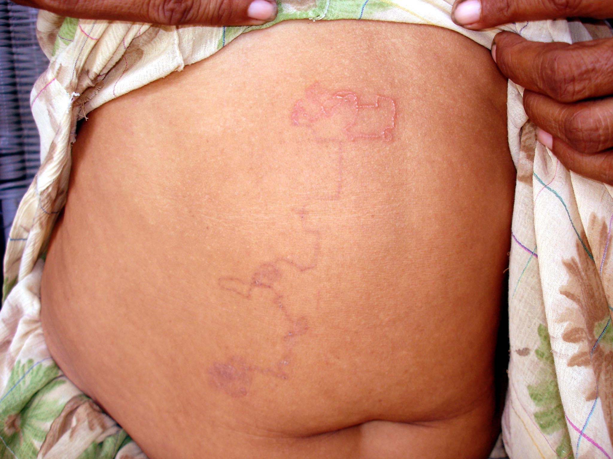
A 50-year-old woman presented with complaints of an itchy eruption on the anterior wall of the abdomen of 2-weeks duration. She was farm worker who spent long hours in fields and she also had pet cats and dogs at her house. She gave no history of fever, cough, dyspnea, or bowel and bladder problem. She was treated with injections of antibiotics and antihistamines with no relief. Cutaneous examination revealed slightly raised, pink, bizarre serpentine-like eruptions with loops and a tortuous path, approximately 35 cm in length over the anterior wall of abdomen extending from lower lumbar region of right side and proceeding upwards to the right hypochondrium. There was healing at the tail end of the lesion.
 |  |
| Figure 1 | Figure 2 |
|---|---|
| Figure 1. Creeping eruption on the anterior abdominal wall | |
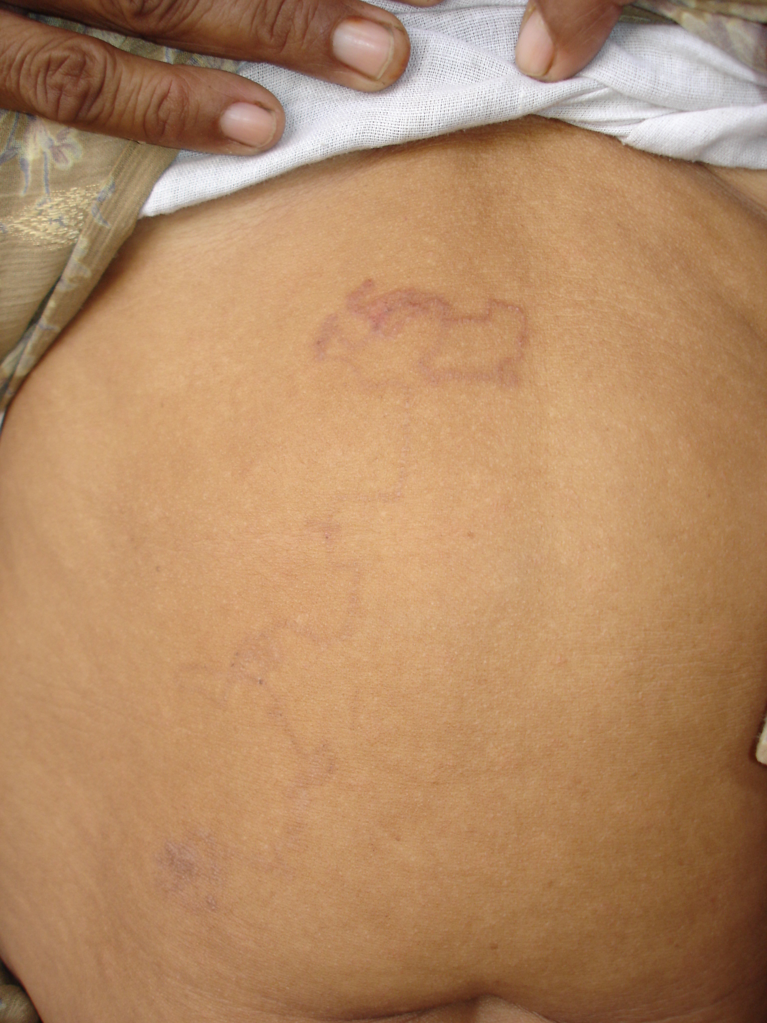
| Figure 2. One week after treatment | |
The baseline laboratory parameters were normal. Biopsy was not done because it is of little value for this condition [2]. Based on the history and clinical findings, a diagnosis of extensive larva migrans was made. Treatment with ivermectin (200 µg/kg body weight) was administered; there was remission after 1 week.
Discussion
Cutaneous larva migrans is also known as sand worms, creeping verminous dermatitis, creeping eruption, plumber's itch, and duck hunter's itch. Numerous organisms are associated with creeping eruption, including Ankylostoma caninum, A. ceylonicum, and A. braziliense, Uncinaria stenocephala, Bubostomum phlebotomum, Gnathostoma spp., Dirofilaria conjunctivae, Capillaria spp., Anatrichostoma cutaneum, Strongyloides stercoralis, Dirofilaria repens, Spirometra spp., Gastrophilus spp., Hypoderma spp., etc. [2].
The most usual form of creeping eruption occurs when the larvae of dog or cat hook worms (Ankylostoma caninum and A. braziliense) penetrate intact, exposed skin and migrate through the epidermis [1]. The most common location is the foot, although other sites including buttocks, back, and thighs (which may have rested on contaminated sand) are susceptible [3]. Reported unusual involvement sites for larva migrans include the penis [4], anterior abdominal wall (5), and oral mucosa and in an infant. Lacking the enzymes necessary to penetrate and survive in the deeper dermis, the larvae wander a serpiginous route at a speed of 3 cm per day. Clinically, the primary lesion is a pruritic, erythematous serpiginous burrow. Although the larvae die usually in 2-8 weeks, survival up to 2 years has been reported. The incubation period ranges from 1 to 6 days. Creeping eruptions are a self-limited dermatosis. Secondary bacterial infection and eczematization are potential complications. Extensive lesions can be associated with wheezing, dry cough, and urticaria. A. caninum larvae can migrate to the small intestine and result in eosinophilic enteritis. Transient eosinophilia is also described [6]. Biopsy is of no value as the larvae advance ahead of the clinical tract. Epiluminescence microscopy is an effective noninvasive method to detect larva and confirm the diagnosis [7].
The lesions disappear in 2-8 weeks but rarely may persist for 2 years. Freezing the leading point of the burrow is an effective older method of treatment. This sometimes produces significant tissue destruction. The larva is up to 2 cm. ahead of the visible burrow; hence, treating the incorrect area will result in treatment failure.
The treatment of choice is ivermectin (a single dose of 200 µg/kg body weight) [8]. Albendazole (400 mg a day by mouth for 3 days) is also effective. An alternative choice of treatment is the topical application of 10 percent topical thiabendazole suspension 4 times a day for at least 2 days after the last sign of burrow activity. Either of the two commercially-available oral preparations may be used directly [9]. This regimen is of great efficacy and has the least toxicity. Rarely, cases are treated with oral thiobendazole.
References
1. Meffert JI. Parasitic infestations. In: Dermatology secrets 1st Ed. Fitzpatrick, Aeling J, editors. New Delhi, India: Jaypee Brothers; 1977. p. 217.2. Vega-Lopez F, Hay RJ. Parasitic worm and protozoa.In: Burns T, Breathnach S, Cox N, Griffiths C, editors. Rooks Textbook of Dermatology. 7th Ed. Oxford; Blackwell; 2004. p. 35.17-8
3. Jelinek T, Maiwald H, Northdurft HD, Loscher T. Cutaneous larva migrans in travelers. Synopsis of histories, symptoms, and treatments of 98 patients. Clin Infect Dis 1994; 19: 1062-6
4. Kartikeyan K, Thappa DM, Jeevankumar B. Cutaneous larva migrans of the penis. Sex Transm Infect 2003; 79:500.
5. Padmavathy L, Rao LL. Cutaneous Larva Migrans - A Case Report. Indian J Med Microbiol [serial online] 2005 [cited 2005 Oct 3]; 23:135-136.
6. Masuria BL, Batra A, Kothiwala RK, Khuller R, Singhi MK. Creeping eruption. Indian J Dermatol Venereol Leprol 1999; 65:51.
7. Eisher E, Themes M, Worret WI. Cutaneous Larva Migrans detected by epiluminescent microscopy. Acta Derm Venereol 1997; 77:487-8.
8. Caumes E, Carriere J, Datry A et al. A randomized trial of ivermectin versus albendazole for treatment of cutaneous larva migrans. Am J Trop Med Hyg 1993; 49: 641-4.
9. Davis CM, Israel RM. Treatment of creeping eruption wih topical thiabendazole. Arch Dermatol 1968; 97:325-6.
© 2006 Dermatology Online Journal