Pseudovitiligo in systemic sclerosis
Published Web Location
https://doi.org/10.5070/D31368x3wxMain Content
Pseudovitiligo in systemic sclerosis
Vandana Mehta Rai MD, and C Balachandran MD
Dermatology Online Journal 11 (3): 41
Department of Skin and STD, KMC Manipal, Karnataka, India
Abstract
Progressive Systemic Sclerosis is a chronic connective tissue disease characterized by sclerosis of skin with involvement of the internal organs. The cause of PSS is unknown but autoimmune mechanisms and primary damage to blood vessels have been proposed to explain its pathogenesis. We report a case of PSS with widespread depigmentation resembling vitiligo.
Progressive Systemic Sclerosis (PSS) is a chronic connective tissue disease characterized by sclerosis of skin with involvement of the internal organs. The cause of PSS is unknown but autoimmune mechanisms and primary damage to blood vessels have been proposed to explain its pathogenesis. We report a case of PSS with widespread depigmentation resembling vitiligo.
|
|
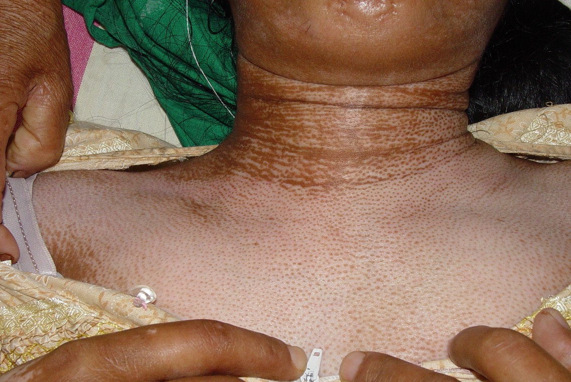
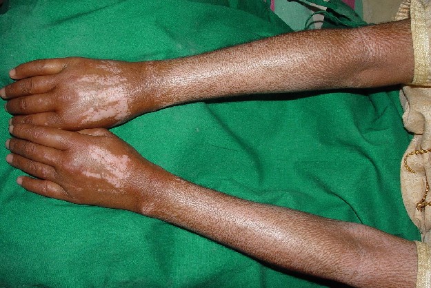
| Figure 1 | Figure 2 |
|---|---|
| Chest (Figure 1) and forearms (Figure 2) | |
A 35-year-old woman presented with diffuse thickening of skin with a history of Raynaud phenomenon, dyspnea, dysphagia, and proximal myopathy. There was no history of photosensitivity, malar rash, or oral ulceration. Cutaneous examination revealed bound-down skin with salt-and-pepper depigmentation affecting the face, forearms, arms, trunk, and lower limbs. Face had a mask-like appearance with a pinched nose, thin pursed lips, and difficulty in eversion of lower eyelids. Widespread depigmentation was the striking feature with perifollicular hyperpigmentation mimicking vitiligo. There was sclerodactyly with digital pitted scars and dilated nail-fold capillaries. Systemic examination was normal. Based on the history and clinical findings a diagnosis of diffuse cutaneous systemic sclerosis was considered. Antibody profile showed a strongly positive result for Scl 70 (++). Skin biopsy was consistent with systemic sclerosis.
figures 3 and 4
Abdomen
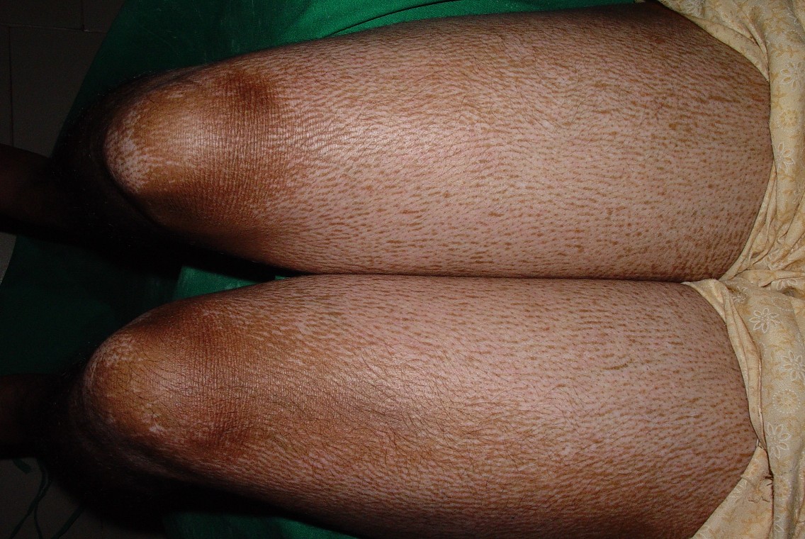
|
| Figure 5 |
|---|
| Thighs |
Changes of hyperpigmentation and depigmentation in PSS have been mentioned in medical literature since 1898[1] but their relationship to the pathogenesis of the underlying condition is poorly understood. The pigmentary alterations seen in PSS are vitiligo-like depigmentation with perifollicular pigment retention, diffuse hyperpigmentation with accentuation in sun exposed areas, and hyperpigmentation and hypopigmentation in areas of sclerosis. The pathogenesis of these lesions is not clear [2]. Vitiligo and the depigmented lesions in PSS share many features in common. Clinically both are characterized by chalk-white macules with well defined borders. Their extensor location possibly suggests some relationship to trauma (Koebner phenomenon) as seen in vitiligo. The extent of involvement, though variable, tends to be more focal and symmetrical in PSS. Mucosal involvement is commonly seen in vitiligo but we have never observed depigmented lesions involving the lips or oral cavity in PSS. Our patient presented with features of PSS with diffuse depigmentation. What was striking was the perifollicular hyperpigmentation mimicking a patch of repigmenting vitiligo. Temperature changes as well as genetic, hormonal, and other physical factors affect pigment formation. Studies show that skin surrounding the hair follicles possesses a richer capillary network that may warm the perifollicular skin and preserve melanogenesis producing the perifollicular pigment retention in PSS [4]. The activation of cellular and humoral immune factors in PSS in combination with external factors such as trauma or inflammation may trigger some series of events that result in the destruction of melanocytes [5].
References
1. Osler WJ. On diffuse scleroderma with special reference to diagnosis and to the use of thyroid gland extract. J Cutan Dis 1998;16:492. Jawitz JC, Albert MK, Nigra TP, Bunning RD. A new skin manifestation of progressive systemic sclerosis. J Am Acad Dermatol 1984;11:265-268
3. Sanchez JL, Vazquez M, Sanchez NP. Vitiligo like macules in Systemic Scleroderma. Arch Dermatol 1983;119:129-133
4. Cronin M, Gerster JC. Pigmentation disorders in Systemic Sclerodermia. Schweiz Rundsch Med Prax 1994;83:42-5
5. Nordlund JJ, Lerner AB, Miller LH. Proceedings of the international workshop on vitiligo. J Invest Dermatol 1978;71:165-166
© 2005 Dermatology Online Journal